Beta Vulgaris Root Tonoplast and Cell Membrane Permeability Increase Through Immersion in Solutions of; Low pH, Extreme Temperature, and of High Solvent Concentration
Eric Horton
March 5th
2012
SBI 4U1 – 70
Prepared for
D. Melegos
Abstract
This
manuscript explores the affects of various stresses on the permeability of cell
membranes and tonoplasts. Some background information will be provided covering
various topics including the fluid mosaic model, free radicals, antioxidants,
betanin, spectrophotometry and Beer’s law. The experimental approach for the
lab will be highlighted along with the objectives and anticipated results. A
clear outline of the methodology followed will be covered followed by the
results obtained through the lab. The results will be explained along with
sources of error present during the lab and future lab prospects.
In order to understand the logic behind
the methodology of this lab, and understand the results, it is important to
have a firm grasp of a few preliminary concepts. For example, the fluid mosaic
model, seen as figure 1 in the
appendix section, is the most widely accepted model used to describe the
composition of biological membranes. The term mosaic implies the nature of the membrane, showing how it is
composed of many different structures that work side by side without creating
an intermediate substance [1]. In general, the structure of
biological membranes consists of a lipid bilayer, with impeded proteins [1].
The term fluid mosaic implies the presence of lateral motion within the membrane.
The proteins float in the lipid bilayer, free to move along the plane of the
membrane [1]. Other components of the membrane include
oligosaccharide chains, membrane proteins and cholesterol [1]. Understanding
the composition of the cell membrane is key to understanding what stresses will
be the most successful in altering the permeability of the Beta vulgaris’ cell
membranes.
Free radicals are very unstable molecules
that possess an odd unpaired electron. This is a result of a bond splitting,
leaving the odd unpaired electron as depicted in figure 2. As a result of their unstable nature, free radicals react
quickly with other compounds attempting to capture the needed electron,
regaining stability. This means that free radicals tend to attack the nearest
stable molecule in order to “steal” an electron, leaving that molecule as a
free radical. This creates a chain reaction that eventually can disrupt the
living cell. Free radicals can be created by your body naturally during
metabolism. Sometimes the body’s immune systems cells purposely create free
radicals in order to neutralize viruses and bacteria present in the body. On
the other hand, environmental factors the body is exposed to such as pollution,
radiation, cigarette smoke and herbicides seen in figure 3 can cause free radical formation. Interestingly enough,
Free radical damage tends to accumulate with age. [2]
Antioxidants
are substances such as vitamin C or E that remove potentially damaging
oxidizing agents from a living organism. Vitamins C and E are thought to
protect the body against the damaging effects of free radicals. These
antioxidants neutralize free radicals by donating one of their own electrons.
This puts an end to the “electron stealing” chain reaction created by free
radicals. Antioxidant nutrients don’t become free radicals themselves after
donating an electron because they are stable in both forms. Their unique
properties help prevent cell and tissue damage that could lead to disease.
Vitamin E tends to protect against cardiovascular disease by defending against
LDL oxidation and artery-clogging plaque formation. Many studies have linked
high vitamin C intakes with lower rates of cancer, particularly cancers of the
mouth, larynx and esophogus. [2]
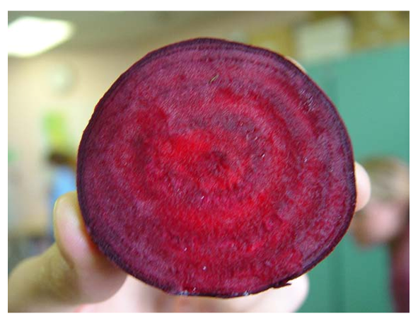
Betanin is a food additive used to give
a red colour to many food products including meat, ice cream and various
beverages. Betanin is found in red beats or Beta
vulgaris and is a type of antioxidant. Being an antioxidant, the
consumption of betanin provides many benefits against oxidative and stress
related disorders. Betanin is also known to increase the red blood cell count
in the human body and maintain a proper metabolism. The flow chart labelled
figure 5 in the appendix section shows the complex series of steps followed
during the biosynthesis of betanin. Betanin has a structure as seen in figure 4, which consists of a great deal of nitrate, which can also be
hazardous to human health in high doses. [3]
Spectrophotometry is the measurement of
a solutions colour by determining the amount of light absorbed in the
ultraviolet, infared, or invisible spectrum, used to calculate the
concentration of a substance in solution [4]. When a beam of
incident light passes through a solution, part of the light is reflected, part
is absorbed and the rest is transmitted. The relationship that exists between
the concentration of a compound and the extent of absorption of light is based
on Lambert’s Law and Beer’s Law [5].
According to Lambert’s law, the light absorbed by a solution is directly
proportional to the length of the light path through the solution [5].
According to Beer’s law, the amount of light absorbed is directly proportional
to the concentration of absorbing solute in the solution [5]. During
this lab, spectrophotometry will be a key tool used to determine the relative
effectiveness of each stress on the Beta vulgaris’ cellular membrane.
The objective of this lab was to
investigate the extracellular concentrations of betanin, and correlate it with
the cell membrane/tonoplast permeability. Factors used to test the viability of
these cell parts will include the determination of the optimal pH, temperature,
and solvent. These results will be quantified using a spectrophotometer.
The approach taken with this experiment
involves various controls coupled with isolation of the various factors. In
order to test for the optimal pH value for cell membrane permeability, five
beet root disks were immersed for 10 minutes in a test tube containing one of
the six solutions, with pH’s of 2,4,6,7,10 and 12. Within this portion of the
experiment, water (pH of 7) acts as a control of sorts. This “neutral” pH
should have little to no effect on the membrane.
For the second portion of the
experiment, temperature was isolated as a factor affecting the membrane
permeability. Five different tests were run, one with five disks inserted into
a test tube with water at a temperature of -180 C, the second set in
a test tube with water at 40 C, the third set was placed in water at
210 C, the fourth set was put in water at 400 C and the
final five disks were put in water at 800 C. All of these solutions
were left to incubate for 10 minutes. The room temperature water in this case
(210 C) acted as a control.
For the final variable, five beet root
disks were put into each of the six test tubes containing various solvents of
varying concentrations for 10 minutes. The solutions used included; 1% acetone,
25% acetone, 50% acetone, 1% methanol, 25% methanol, 50% methanol.
After the incubation time for each of
the samples was complete, the extracellular fluid from each test tube was
poured into a small cuvette and then inserted into the spectrophotometer. The
resulting absorbance values were examined and a conclusion was drawn.
It is anticipated that the greatest
absorbance will occur when the cells have been submitted to the harsher
stresses. Various high and very low temperatures should return a high
absorbance value, along with the higher concentrations of solvents. High and
low pH’s should also cause greater cell membrane and tonoplast permeability,
resulting
in higher absorbance values. The more moderate conditions aren’t as likely to
increase the cells permeability and in turn should have low absorbance values.
Methodology
The methodology for this lab was broken
down into several steps. The first step involved the preparation of the Beta
vulgaris root for testing. The beet was prepared by slicing the top and bottom
off of a fresh beet. Using a cork borer, a core sample of the root was
extracted. After driving the borer into the beet, the tweezers were used to remove
the core sample. The cylindrical root section was then sliced into small 3mm
thick disks using a scalpel. For our section of the experiment, fifteen disks
were prepared. Once all the cutting was complete, the disks were put into a
beaker and then immersed in tap water. The beaker was then agitated, swirled
and then emptied. This process was repeated three times in order to adequately
rinse the contents of the burst cells from the samples.
In order to test the permeability of the
cells under various temperature conditions, five pre cut and rinsed disks were
placed in the freezer at a temperature of eighteen degrees below zero Celsius.
Five disks were also placed in the fridge at a temperature of four degrees
Celsius. Five disks from the freezer were then put into a large test tube, five
disks from the fridge were placed in another large test tube, and then the five
of freshly cut disks were placed into each of the three remaining test tubes. Using
a graduated cylinder, 10ml of tap water was measured out and added to each of
the five test tubes. Each of the samples was incubated for 10 minutes. The
samples containing the disks from the fridge and freezer were incubated at room
temperature along with the 210 C sample. The 400C and 800C
samples were incubated in water baths at their assigned temperatures.
In order to investigate the relationship
between membrane and tonoplast permeability and varying pH, a series of steps
were followed. As for the previous test, 25 3mm beet root disks were prepared
using the cork borer and scalpel. 5 disks were placed into each of the six
clean test tubes prepared. Each test tube was labelled using masking tape and a
permanent marker with the pH value of either 2,4,6,7,10 or 12. Then 10mL of
each solution of a varying pH was measured using a graduated cylinder, and then
added to the corresponding test tube.
As for the test involving pH, a key
sequence of steps was followed in order to test the effect of various solvents
of numerous concentrations on membrane and tonoplast permeability. 25 3mm beat
root disks were prepared using a cork borer and a scalpel. 5 disks were placed
in each of the 6 test tubes. Each test tube was labelled with one of the
following solvents and concentration; 1% acetone, 25% acetone, 50% acetone 1%
methanol, 25% methanol, 50% methanol. Then 10ml of each solvent was measured
using a graduated cylinder and added to the corresponding test tube.
All of the samples were then allowed to
incubate for 10 minutes, some at specific temperatures, and the rest at room
temperature. After the 10 minute incubation period, the samples were gently
agitated and then the liquid portion was poured into small test tubes. The
spectrophotometer was then set to 560nm and calibrated using a test tube
containing tap water. Once calibrated, each of the five small test tubes were
placed in the spectrophotometer and their absorbance readings were recorded in table 1 in the appendix section.
Results
After
conducting the above experiment, a clear and relatively definitive set or
results were obtained. As seen in table
1, it was clearly observed that as the different stresses were applied,
varying absorbance values resulted. With regards to pH, the lower acidic pH
values returned higher absorbance values than the moderate to basic pH
solutions. At a pH of 2, the highest absorbance value of 0.52 was observed
compared to the 0.02 value at the pH of water and 0.05 at a pH of 12. This
clearly indicates that the cell membranes and tonoplasts are significantly more
permeable in acidic solutions. As indicated by chart 1 found in the appendix section, a clear logarithmic trend
associating increasing pH with reduced absorbance values can be observed.
The temperature related stress
results were rather interesting as well. Table
1 indicates that the cell membrane and tonoplast are the most permeable at
extreme temperatures. At -180 C an absorbance value of 1.61 is
observed and at 800 C an absorbance value of 1.05 results. These
values contrast the absorbencies indicated at a more moderate temperature of 210
C where absorbance values of 0.04 were observed. Through plotting this
data on chart 2, a parabolic trend
is observed, this indicates that extreme temperatures provide the most stress
to a cells membrane and tonoplasts.
In the final test, it was noted that
with increasing the concentration of solvent used in the test, greater membrane
and tonoplast permeability was achieved. The absorbencies for this test ranged
from 0.02 for the 1% acetone solution, to 0.46 for the 50% acetone solution. For
the methanol, 0.02 absorbance values was observed at a 1% concentration, and at
a 50% concentration a 0.32 absorbance value was found to exist. Through the
exponential trends illustrated on chart
3 it became clear that not only as the concentration increased the
absorbency increased, but the acetone was more effective than methanol at
creating permeability.
Discussion
The
results obtained through the series of test conducted can be justified using
some basic knowledge of cell membranes. The results of the temperature portion
of the lab can be explained using the properties of cell membranes. When the
cell is submitted to high temperatures, the phospholipids become increasingly
fluid, increasing the permeability of the membrane. This allows materials
previously unable to travel in and out of the cell to do so. The extremely high
temperatures can also break the hydrogen bonds found in the protein structure
which could cause protein de-naturation. When the cell is frozen, the membrane
bursts because of the ice crystals and extreme temperatures make the cell
brittle, causing it to fracture. This allows an increase in permeability as
well.
When subjected to a solution consisting
of a solvent the membrane also becomes more permeable. Acetone and methanol are
both polar solvents. The phospholipids bilayer is also polar, meaning they will
tend to combine easily. Solvents denature the proteins found in the
phospholipids bilayer, meaning the sulphide bridges between cysteine amino
acids found in the quaternary and tertiary may be disrupted. The alteration to
the proteins structure stops them from being able to function correctly. This
protein alteration creates gaps in the cell membrane that allow for increased
permeability. As the concentration of the solvents increased, this effect is
further magnified. Acetone has a greater effect on the cell membrane because
acetone has a greater molecular dipole (2.91) than that of methanol (1.69). The
difference in polarity makes acetone a stronger solvent, resulting in a more
significant effect on the cell membrane.
pH also has a similar affect on the cell
membrane. With the increase in acidity during the pH stress test, protein
denaturation occurs as well. The change in pH tends to disrupt the ionic bonds
found in the proteins tertiary and quaternary structures. These disruption
cause changes to the shape of the protein and as a result of this the protein
is denatured. Changes to the proteins composition can cause an increase in the
membrane permeability.
Along
with the increase in permeability through each of the stresses, comes an
increase in the absorbency. As the cell membrane becomes increasingly
permeable, the more betanin is released into the extracellular fluid. With the
increase in betanin release, there is an increase in the absorbency measured
using the spectrophotometer.
Through conducting this lab several
sources of error became evident. During the temperature stress test, the
incubation time was supposed to be 10 minutes. However, by the time the water
was added to each of the test tubes, and each test tube made it to its
designated temperature environment, the times were skewed. With the present lab
set up it was impossible to incubate each of the samples for the same period of
time. The difference in incubation time could alter the absorbency values
significantly. This could be remedied by setting separate timers for each of
the test tubes incubation. Another source of error that became evident was the
insertion of the frozen or chilled beet disks into room temperature water. This
would moderate the temperature during incubation, rather than having a
consistent temperature. This problem however is somewhat unavoidable, unless
that water was made the same temp as the disk. This would pose a problem for
the frozen disk test however. The final source of error that was evident during
the lab was the consistency in the size of beet disks used. There was no step
taken to ensure absolute accuracy with respect to disk size. Different disk
sizes could contribute to large or fewer available cells and therefore lesser
or greater absorbency values. This problem could be fixed using a tool that
cuts consistent disk sizes. An example of this may include a cheese slicer. A
consistent disk size would make for far more accurate results.
A potential lab proposal may be one that
incorporates a comparison between polar and non polar solvents. Despite its
carcinogenic nature, benzene would be a good non polar solvent that could be
compared to acetone and methanol. 3 test tubes, one with 1% concentration the
others 25% and 50% respectively. The results of this test could be graphed with
the results of the polar solvent test. This would give insight into the effects
of the disruption of the London forces between non polar amino acids. Another
potential lab proposal might involve the isolation of betanin in order to
explore its abilities as an antioxidant. One might consider introducing free
radicals into a group of plant or animals and observing the effects. Then a
comparison might be made with the results after antioxidants are added to the
samples. This would allow us to verify and explore the importance of
antioxidants in our diet.
References
[1] Campbell, M. K., & Farrell, S. O. (2011).
Lipids and Proteins are Associated in Biological Membranes. Biochemistry
(7th ed., pp. 207,208). Belmont: Brooks/Cole.
[5] Chauhan, B. (2008). Principles of Biochemistry
and Biophysics. Bangalore, New Delhi: University Science Press.
[2] Understanding Free Radicals and Antioxidants.
(n.d.). HealthCheck Systems. Retrieved March 3, 2012, from
http://www.healthchecksystems.com/antioxid.htm
[3] Walker, M. (2011, February 25). What is Betanin
Used For? Health Benefits and Side Effects of Betanin. Kay Circle | Everyday
Reference. Retrieved March 3, 2012, from http://www.kaycircle.com/What-is-Betanin-Used-For-Health-Benefits-and-Side-Effects-of-Betanin
[4] spectrophotometry - definition of
spectrophotometry in the Medical dictionary - by the Free Online Medical
Dictionary, Thesaurus and Encyclopedia.. (n.d.). Medical Dictionary.
Retrieved March 3, 2012, from
http://medical-dictionary.thefreedictionary.com/spectrophotometry


